Anorectal Disorders – Fissure, Fistula, Hemorrhoids, Pilonidal Sinus & Abscess service image
Anorectal Disorders – Fissure, Fistula, Hemorrhoids, Pilonidal Sinus & Abscess

Anorectal disorders affect the anus, rectum and surrounding tissues and can cause pain, bleeding, swelling, itching, discharge and difficulty with bowel movements. Common conditions include anal fissure, anal fistula, hemorrhoids, perianal abscess and pilonidal sinus. Dr. Shaunak Saha provides evaluation and treatment planning for patients experiencing anorectal symptoms. A detailed clinical assessment helps identify the underlying condition and determine whether conservative treatment, medication, drainage, minimally invasive treatment or surgery is appropriate. An anal fissure is a small tear in the anal lining and can cause sharp pain and bright-red bleeding during or after bowel movements. Many fissures improve with conservative treatment, while persistent or recurrent cases may require a procedure. An anal fistula is an abnormal tunnel between the anal canal and nearby skin, often developing after an anal abscess. Fistulas generally do not heal on their own and may require surgical treatment. The choice of procedure depends on the fistula's location and relationship to the anal sphincter muscles. Treatment for anorectal conditions is planned according to the diagnosis, severity, symptoms and individual patient factors, with attention to preserving normal bowel function wherever possible. SPECIFICATIONS Conditions: Anal fissure, anal fistula, hemorrhoids and perianal abscess Additional condition: Pilonidal sinus Assessment: Clinical anorectal examination Investigations: Proctoscopy, ultrasound, MRI or other tests when clinically indicated Treatment options: Medical, conservative, minimally invasive or surgical Fistula treatment: Fistulotomy, seton or other appropriate procedures depending on anatomy Fissure treatment: Medical management and procedures for persistent or recurrent disease Abscess treatment: Drainage when clinically required Hemorrhoid treatment: Non-surgical, minimally invasive or surgical treatment according to severity Treatment planning: Individualised colorectal assessment FREQUENTLY ASKED QUESTIONS Q: What are common anorectal disorders? A: Common conditions include anal fissure, anal fistula, hemorrhoids, perianal abscess and pilonidal disease. Q: What is the difference between a fissure and a fistula? A: An anal fissure is a tear in the anal lining, while an anal fistula is an abnormal tunnel connecting the anal canal with the surrounding skin. Q: Does an anal fistula require surgery? A: Most anal fistulas do require a surgical procedure because they usually do not heal on their own. The appropriate procedure depends on the fistula's location and whether it involves the anal sphincter muscles. Q: What are symptoms of an anal abscess? A: An anal abscess can cause painful swelling, redness and sometimes fever. It may require prompt medical assessment and drainage. Q: Can anal fissures be treated without surgery? A: Yes. Many anal fissures improve with measures such as keeping stools soft, dietary fibre, fluids and appropriate medical treatment. Persistent or recurrent fissures may require a procedure. Q: When should I see a colorectal surgeon? A: Persistent anal pain, recurrent swelling, discharge, bleeding, a suspected fistula or symptoms that do not improve with initial treatment should be evaluated by a qualified doctor. Don't ignore persistent anal pain, bleeding, swelling or discharge. Consult Dr. Shaunak Saha for an accurate diagnosis and personalised treatment plan for anorectal conditions.

Open Hernia Repair service image
Open Hernia Repair

Open hernia repair is a surgical procedure used to treat a hernia through an incision made directly over or near the affected area. A hernia occurs when tissue from inside the abdomen pushes through a weakened area of the abdominal wall, often causing a visible or palpable bulge and discomfort. Dr. Shaunak Saha provides evaluation and surgical treatment for patients with inguinal and other abdominal wall hernias. During consultation, the type, size and location of the hernia, symptoms, previous surgeries and overall health are assessed before selecting the appropriate surgical approach. During open hernia repair, the protruding tissue is returned to its appropriate position and the weakened area of the abdominal wall is repaired. Mesh reinforcement may be used when clinically appropriate to strengthen the repair and reduce the risk of recurrence. Open surgery may be particularly appropriate for certain larger, recurrent or complicated hernias and in situations where laparoscopic surgery is not the preferred approach. The choice between open and laparoscopic repair is made according to the individual patient's condition. SPECIFICATIONS Procedure: Open hernia repair Common conditions: Inguinal and selected abdominal wall hernias Surgical approach: Open surgery Assessment: Clinical examination and imaging when required Repair: Reduction of hernia contents and repair of the abdominal wall defect Mesh: May be used when clinically indicated Anaesthesia: Local, regional or general depending on the case Treatment planning: Individualised according to hernia type and patient health Recovery: Depends on the hernia, surgical technique and individual factors Follow-up: Postoperative wound and surgical review FREQUENTLY ASKED QUESTIONS Q: What is open hernia repair? A: Open hernia repair is an operation in which the surgeon accesses the hernia through an incision, returns the protruding tissue to its appropriate position and repairs the weakened abdominal wall. Q: Is mesh always required for open hernia surgery? A: No. Mesh is commonly used for many types of hernia repair, but whether mesh is appropriate depends on the type and characteristics of the hernia and the surgical technique. Q: Is open or laparoscopic hernia surgery better? A: Neither approach is universally better. The appropriate technique depends on the type and size of the hernia, previous surgery, overall health and other individual factors. Q: Can a hernia become an emergency? A: A hernia can occasionally become trapped and severely painful, which may require urgent surgical treatment. Sudden severe pain, a hard or irreducible swelling, vomiting or other concerning symptoms require urgent medical assessment. Have a persistent groin or abdominal swelling? Consult Dr. Shaunak Saha for a detailed hernia evaluation and discuss whether open hernia repair or another treatment approach is appropriate for you.

Varicose Veins Surgery service image
Varicose Veins Surgery

Varicose veins are enlarged, twisted veins that most commonly occur in the legs. They can be associated with aching, swelling, heaviness and visible changes in the veins. Evaluation may include physical examination and venous Doppler ultrasound to assess blood flow and the condition of the veins. Dr Shaunak Saha provides consultation and treatment planning for patients with symptomatic or clinically significant varicose veins. Treatment depends on the severity of the condition and may include lifestyle measures, compression therapy, minimally invasive procedures or surgery. Available treatments for varicose veins can include sclerotherapy, laser-based treatment, radiofrequency or laser endovenous ablation, phlebectomy and, in selected cases, conventional vein surgery. SPECIFICATIONS: Condition: Varicose veins Affected area: Lower limbs Symptoms: Pain, aching, swelling or heaviness Assessment: Clinical examination Imaging: Venous Doppler ultrasound when indicated Treatment: Conservative, minimally invasive or surgical Procedures: Ablation, sclerotherapy, phlebectomy or surgery as appropriate Treatment setting: Often outpatient for selected procedures Goal: Symptom management and treatment of affected veins Planning: Individualised FREQUENTLY ASKED QUESTIONS: Q: What are varicose veins? A: Varicose veins are enlarged and twisted veins, usually visible in the legs. Q: Do all varicose veins need surgery? A: No. Treatment depends on symptoms, severity and the underlying venous problem. Options can include compression, lifestyle measures and procedures. Q: What test is commonly used to assess varicose veins? A: Venous Doppler ultrasound can evaluate blood flow and help assess the veins. If varicose veins are causing pain, swelling, heaviness or concern about their appearance, consult Dr Shaunak Saha for evaluation and treatment options.

Pediatric Hernia Treatment and Surgery service image
Pediatric Hernia Treatment and Surgery

Pediatric hernia is a common surgical condition in which tissue from inside the abdomen can push through a weak or incompletely closed area, often producing a groin or abdominal bulge. In children, inguinal hernias are commonly related to developmental factors. Dr Shaunak Saha provides consultation and surgical evaluation for children with suspected hernia. Treatment is planned according to the child’s age, type of hernia, symptoms and clinical findings. Pediatric hernia repair may be performed using an open approach or laparoscopic keyhole surgery depending on the individual case. Children undergoing surgery are generally assessed carefully by the surgical and anaesthesia teams before treatment. SPECIFICATIONS: Condition: Pediatric hernia Common type: Inguinal hernia Patient group: Infants and children Assessment: Pediatric clinical examination Treatment: Surgical repair when indicated Approach: Open or laparoscopic Anaesthesia: Usually general anaesthesia Recovery: Often rapid in uncomplicated cases Follow-up: Postoperative review Parent support: Preoperative and postoperative guidance FREQUENTLY ASKED QUESTIONS: Q: What is a pediatric hernia? A: It is a hernia occurring in an infant or child, commonly presenting as a groin or abdominal bulge. Q: Does a child's hernia need surgery? A: Pediatric inguinal hernias generally require surgical repair because they do not usually close on their own and can carry a risk of complications. Q: Can pediatric hernia surgery be done laparoscopically? A: Yes. Laparoscopic keyhole repair may be an option for selected children. If your child has a groin or abdominal swelling that may be a hernia, consult Dr Shaunak Saha for timely pediatric surgical assessment.

Circumcision – Pediatric and Adult service image
Circumcision – Pediatric and Adult

Circumcision is a surgical procedure that removes the foreskin covering the head of the penis. It may be performed for medical, cultural, religious or personal reasons. Different techniques may be used depending on the patient’s age, anatomy, indication and surgeon’s assessment. Dr Shaunak Saha provides consultation and surgical care for selected pediatric and adult circumcision cases. The procedure may involve conventional open techniques or device-assisted/stapler techniques where appropriate and available. Before circumcision, the patient or parents should receive clear information about the reason for the procedure, available techniques, expected recovery and potential risks. SPECIFICATIONS: Procedure: Circumcision Patient groups: Pediatric and adult Indications: Medical, cultural, religious or personal Techniques: Open or device-assisted techniques where appropriate Assessment: Clinical examination Anaesthesia: Depends on age and procedure Treatment setting: Hospital or appropriate surgical facility Recovery: Depends on age and technique Aftercare: Wound care and activity guidance Follow-up: As advised FREQUENTLY ASKED QUESTIONS: Q: What is circumcision? A: Circumcision is the surgical removal of the foreskin covering the head of the penis. Q: Is circumcision performed in adults as well as children? A: Yes. Circumcision can be performed in newborns, children and adults depending on the circumstances. Q: How long does circumcision take to heal? A: Healing varies by age and technique. Patients should follow the specific postoperative instructions provided by their doctor. For pediatric or adult circumcision consultation, speak with Dr Shaunak Saha about the appropriate technique and treatment plan.

Parotid Swellings Diagnosis and Surgical Treatment service image
Parotid Swellings Diagnosis and Surgical Treatment

The parotid glands are major salivary glands located in front of the ears. A swelling or lump in the parotid region can have several causes, including benign and malignant tumours. Many parotid tumours are benign, but proper evaluation is important because some can be cancerous. Dr Shaunak Saha provides clinical assessment and surgical treatment planning for patients presenting with parotid swellings. Evaluation may involve physical examination, imaging and tissue assessment depending on the nature of the swelling. Surgical planning for parotid disease requires careful consideration of the facial nerve and surrounding structures. The recommended treatment depends on the diagnosis, size, location and characteristics of the swelling. SPECIFICATIONS: Condition: Parotid swelling Anatomical area: Parotid salivary gland Assessment: Clinical examination Imaging: Ultrasound or other imaging when indicated Tissue diagnosis: Biopsy or sampling when appropriate Treatment: Observation or surgery depending on diagnosis Surgical consideration: Facial nerve preservation Target: Benign and selected malignant parotid lesions Planning: Individualised Follow-up: Based on pathology and treatment FREQUENTLY ASKED QUESTIONS: Q: What causes a parotid swelling? A: Parotid swellings can have several causes, including benign and malignant tumours. Q: Are parotid tumours cancerous? A: Many parotid tumours are benign, but some are malignant, which is why evaluation is important. Q: Why is facial nerve protection important during parotid surgery? A: The facial nerve passes through the parotid gland, so careful surgical planning is important to protect facial movement. If you notice a persistent swelling or lump near the ear or jaw, consult Dr Shaunak Saha for appropriate parotid evaluation.

Breast and Thyroid Swellings Evaluation and Surgical Treatment service image
Breast and Thyroid Swellings Evaluation and Surgical Treatment

Breast and thyroid swellings can have many causes, ranging from benign conditions to conditions requiring further investigation or treatment. A breast lump may be caused by cysts, fibroadenoma, fibrocystic changes, infection or other conditions, while thyroid nodules may be solid or fluid-filled and are often benign. Dr Shaunak Saha provides clinical assessment and treatment planning for patients presenting with breast lumps, thyroid swellings and related concerns. Depending on the clinical findings, evaluation may include examination, ultrasound, thyroid function tests, imaging and tissue sampling such as fine-needle aspiration when appropriate. The purpose of evaluation is to identify the nature of the swelling and determine whether observation, further investigation or surgical treatment is appropriate. SPECIFICATIONS: Conditions: Breast and thyroid swellings Assessment: Clinical examination Breast evaluation: Imaging and tissue assessment when indicated Thyroid evaluation: Thyroid function testing and ultrasound Biopsy: Fine-needle aspiration when clinically appropriate Treatment: Observation, investigation or surgery Surgical focus: Removal or treatment of selected lesions Patient group: Adults requiring breast or thyroid evaluation Follow-up: Based on diagnosis Planning: Individualised FREQUENTLY ASKED QUESTIONS: Q: Should every breast lump be checked? A: Yes. Many breast lumps are benign, but a new or unusual breast lump should be medically assessed. Q: Are thyroid nodules usually cancerous? A: Most thyroid nodules are not cancerous, but appropriate evaluation is important to determine their nature. Q: What tests may be needed for a thyroid swelling? A: Depending on the case, evaluation can include thyroid blood tests, ultrasound and fine-needle aspiration biopsy. Do not ignore a new breast or thyroid swelling. Book a consultation with Dr Shaunak Saha for appropriate clinical evaluation.

Lumps and Bumps Treatment service image
Lumps and Bumps Treatment

Lumps and bumps can have many causes, including sebaceous or epidermoid cysts, lipomas, ganglion cysts and other benign or more serious conditions. A new or changing lump should be assessed rather than assumed to be harmless. Dr Shaunak Saha provides clinical evaluation of common soft-tissue lumps and swellings and can advise whether observation, imaging, biopsy or surgical removal may be appropriate. The treatment approach depends on the location, size, symptoms, appearance and clinical characteristics of the lump. Where surgical removal is recommended, the aim is to treat the identified lesion while considering the surrounding tissue and cosmetic outcome. SPECIFICATIONS: Conditions: Cysts, lipomas, ganglion cysts and selected lumps Assessment: Clinical examination Additional evaluation: Imaging or biopsy when indicated Treatment: Observation or surgical removal Procedure: Excision where appropriate Target area: Skin and soft tissues Anaesthesia: Depends on location and procedure Pathology: Tissue may be sent for laboratory examination Recurrence: Depends on lesion type Follow-up: Based on diagnosis and treatment FREQUENTLY ASKED QUESTIONS: Q: Are all lumps and bumps harmless? A: No. Many are benign, but a new, growing, painful or otherwise unusual lump should be evaluated. Q: Can a lipoma or cyst be removed surgically? A: Yes, when clinically appropriate, symptomatic or otherwise requiring removal. Q: Will every lump require a biopsy? A: No. The need for imaging, biopsy or removal depends on the clinical characteristics of the lump. If you have a new, persistent or growing lump or swelling, consult Dr Shaunak Saha for proper evaluation before deciding on treatment.

Pilonidal Sinus Treatment and Surgery service image
Pilonidal Sinus Treatment and Surgery

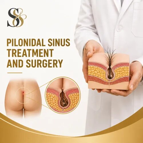
Pilonidal sinus is a condition affecting the cleft between the buttocks and can cause a painful swelling, drainage or recurrent infection. Treatment depends on the severity and whether the condition is recurring. Dr Shaunak Saha provides assessment and treatment planning for patients with pilonidal sinus and recurrent pilonidal disease. A healthcare professional can often diagnose the condition through medical history and physical examination. Treatment can range from drainage of an abscess to surgical removal of the affected sinus when appropriate. Wound management and strategies to reduce recurrence are important parts of care. SPECIFICATIONS: Condition: Pilonidal sinus Location: Natal cleft Common symptoms: Pain, swelling and drainage Assessment: Clinical examination Treatment: Drainage or surgery depending on condition Surgical option: Excision of affected sinus Anaesthesia: Local or general depending on procedure Recurrence: Requires preventive planning Aftercare: Wound care and follow-up Suitability: Individualised FREQUENTLY ASKED QUESTIONS: Q: What is pilonidal sinus? A: It is a condition involving a sinus or cyst in the cleft between the buttocks, which can become infected. Q: Does pilonidal sinus always require surgery? A: Not always. An acute abscess may first require drainage, while recurrent disease may require surgical treatment. Q: Can pilonidal sinus come back after surgery? A: Recurrence is possible, and the risk varies depending on the disease and surgical approach. For persistent, painful or recurrent pilonidal sinus, consult Dr Shaunak Saha for diagnosis and treatment planning

Hydrocele Treatment and Surgery service image
Hydrocele Treatment and Surgery

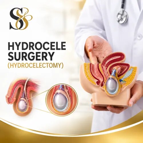
A hydrocele is a collection of fluid around a testicle that can cause swelling of the scrotum. Hydroceles are common in newborns and can also occur in older children and adults. Many hydroceles are painless, but persistent or significant swelling should be medically evaluated. Dr Shaunak Saha provides clinical assessment for patients with hydrocele and other causes of scrotal swelling. Evaluation may include physical examination and, when required, ultrasound to distinguish hydrocele from hernia, tumour or other conditions. If a hydrocele does not resolve or causes significant symptoms, surgical repair may be recommended. The appropriate treatment depends on the patient's age, symptoms, underlying cause and clinical findings. SPECIFICATIONS: Condition: Hydrocele Affected area: Scrotum Cause: Fluid collection around the testicle Assessment: Physical examination Imaging: Ultrasound when required Treatment: Observation or surgery depending on case Procedure: Hydrocele repair Anaesthesia: Based on age and procedure Follow-up: Postoperative review where surgery is performed Suitability: Individualised FREQUENTLY ASKED QUESTIONS: Q: What is a hydrocele? A: A hydrocele is a collection of fluid around a testicle that can cause scrotal swelling. Q: Does every hydrocele need surgery? A: No. Some hydroceles, particularly in infants, may resolve without treatment. Persistent or symptomatic hydroceles may require surgery. Q: Can hydrocele be confused with a hernia? A: Yes. A clinical examination and sometimes ultrasound may be needed to distinguish hydrocele from hernia or other causes of scrotal swelling. If you notice persistent scrotal swelling, consult Dr Shaunak Saha for evaluation and an appropriate treatment plan.

Laparoscopic Liver Abscess Drainage and Treatment service image
Laparoscopic Liver Abscess Drainage and Treatment

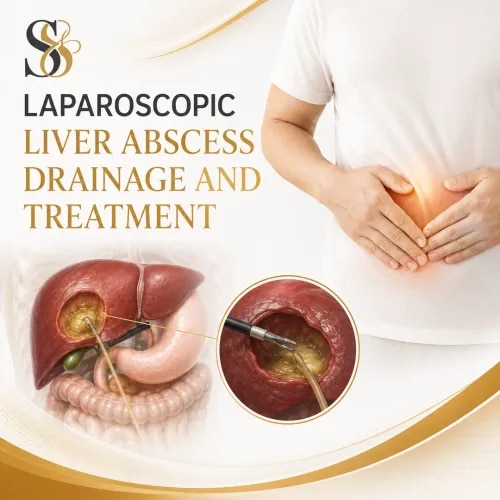
A liver abscess is a collection of infected fluid or pus within the liver. Diagnosis generally involves clinical assessment and imaging such as ultrasound or CT, while treatment may involve antibiotics and drainage depending on the type, size, location and severity of the abscess. Dr Shaunak Saha provides surgical evaluation for patients with suspected or diagnosed liver abscesses requiring procedural or surgical management. When drainage is necessary, the appropriate method is selected according to the abscess location and the patient’s condition. Liver abscesses require timely medical attention because untreated abdominal abscesses can result in serious complications, including systemic infection. SPECIFICATIONS: Condition: Liver abscess Treatment: Drainage and medical management Assessment: Clinical examination and imaging Imaging: Ultrasound or CT where appropriate Approach: Minimally invasive or surgical drainage depending on case Additional treatment: Antibiotics when clinically indicated Target organ: Liver Treatment planning: Individualised Monitoring: Clinical and imaging follow-up Urgency: Requires timely medical assessment FREQUENTLY ASKED QUESTIONS: Q: What is a liver abscess? A: It is a collection of infected fluid or pus within the liver. Q: How is a liver abscess treated? A: Treatment can include antibiotics and drainage, depending on the type and characteristics of the abscess. Q: Is a liver abscess serious? A: It can become serious if untreated, so prompt medical evaluation is important. If you have been diagnosed with a liver abscess or have symptoms requiring urgent evaluation, consult Dr Shaunak Saha for appropriate assessment and treatment planning.

Laparoscopic Splenectomy service image
Laparoscopic Splenectomy

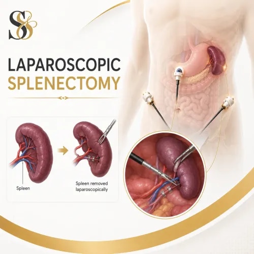
aparoscopic splenectomy is a minimally invasive surgical procedure to remove the spleen through small abdominal incisions. Splenectomy may be required for selected conditions involving the spleen, including certain blood disorders, trauma, enlargement, infections or other diseases. Dr Shaunak Saha provides surgical evaluation and treatment planning for patients who may require spleen surgery. The decision to remove the spleen is based on the underlying condition, severity, symptoms, test results and overall health. Because the spleen contributes to immune function, patients who undergo splenectomy require appropriate medical planning and preventive care, including vaccination and infection-prevention advice as recommended by their healthcare team. SPECIFICATIONS: Procedure: Laparoscopic splenectomy Target organ: Spleen Approach: Minimally invasive laparoscopic surgery Indications: Selected spleen disorders Assessment: Imaging, blood tests and clinical evaluation Anaesthesia: Usually general anaesthesia Incisions: Small abdominal incisions Treatment planning: Individualised Postoperative care: Infection-prevention planning Follow-up: Required after surgery FREQUENTLY ASKED QUESTIONS: Q: Why might someone need a splenectomy? A: Reasons can include certain blood disorders, traumatic spleen injury, enlarged spleen, some cancers and selected infections. Q: Can a person live without a spleen? A: Yes, but people without a spleen have an increased risk of certain serious infections and need appropriate preventive healthcare. Q: Is laparoscopic splenectomy suitable for everyone? A: No. The surgical approach depends on the spleen condition, anatomy, previous surgery and overall health. For evaluation of a spleen condition requiring surgical treatment, consult Dr Shaunak Saha to discuss the appropriate treatment approach.

Subcategory Tags

Abdominal AbscessAbdominal HerniaAbscess DrainageAcute Abdominal PainAdult CircumcisionAnal Fissure SurgeryAnal Fissure TreatmentAnal Fistula SurgeryAnal Fistula TreatmentAnorectal Disorders,Anorectal SurgeryAppendicitis SurgeonAppendicitis TreatmentAppendix RemovalAppendix SurgeryBaby HerniaBreast Lump Evaluation,Breast Lump TreatmentBreast SurgeryBreast and Thyroid SurgeryChild Hernia SurgeryCircumcisionCircumcision SurgeryColorectal SurgeryCyst RemovalEndovenous TreatmentFacial Nerve SurgeryForeskin SurgeryGallbladder RemovalGallbladder SpecialistGallbladder SurgeryGallstone SurgeonGallstone TreatmentGanglion CystGeneral SurgeonGeneral SurgeryGroin HerniaGynaecological SurgeryHead and Neck SurgeryHemorrhoids TreatmentHepatobiliary SurgeryHernia SurgeryHernia TreatmentHernia Treatment PuneHydrocele RepairHydrocele SurgeryHydrocele TreatmentInguinal HerniaInguinal Hernia SurgeryInguinal Hernia in ChildrenKeyhole Hernia SurgeryKeyhole SurgeryLaparoscopic AppendectomyLaparoscopic CholecystectomyLaparoscopic Colorectal SurgeryLaparoscopic Hernia RepairLaparoscopic HysterectomyLaparoscopic Pediatric SurgeryLaparoscopic RectopexyLaparoscopic Splenectomy

Contact Us

Live Chat

Connect over WhatsApp to resolve your queries realtime.