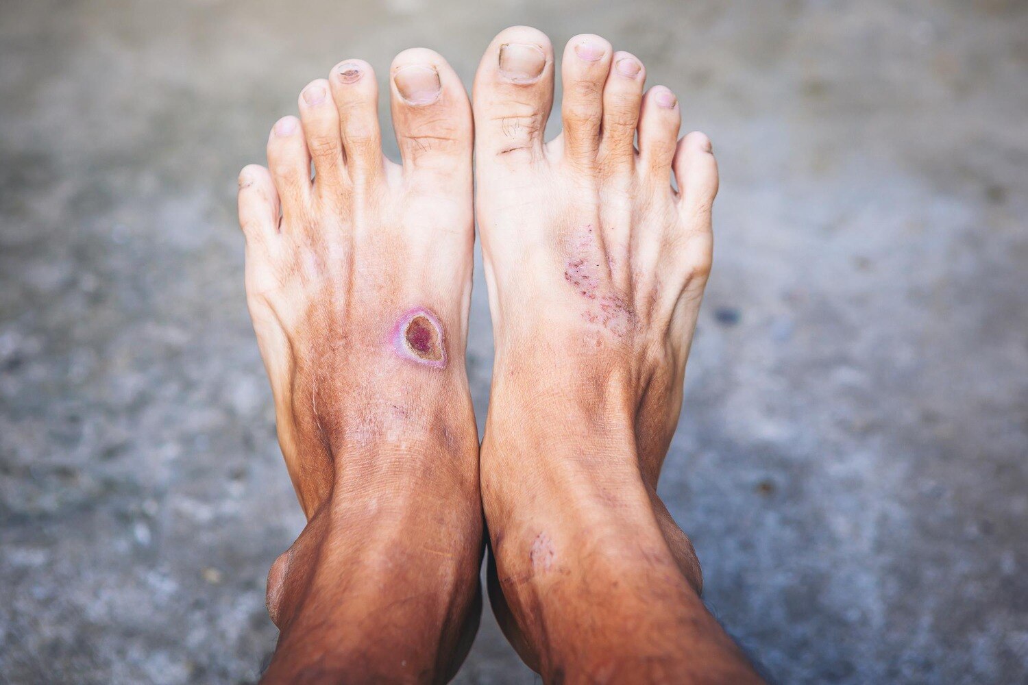
A blister, crack, burn, or pressure mark on a diabetic foot can worsen beneath apparently minor surface damage. By the end, you will know why pain is an unreliable warning, which changes require same-day care, what an evaluation checks, and how circulation, infection, depth, and pressure determine treatment.
Key takeaways
- Have every diabetic foot wound examined, even if it does not hurt.
- Seek same-day care for spreading redness, swelling, pus, fever, or blackened skin.
- X-rays, blood tests, and circulation checks can reveal deeper complications.
- Keep pressure off the wound and follow prescribed cleaning and treatment instructions.
Why a small or painless wound can become dangerous
Do not wait for pain before having a diabetic foot wound examined. Diabetic peripheral neuropathy can cause protective sensation loss, so you may not feel pressure, heat, a foreign object, or a wound becoming deeper.
A small diabetic foot wound from shoe friction can repeatedly reopen with every step, especially over a toe, heel, or callus.
Dryness does not prove safety. A painless diabetic foot ulcer can extend into deeper tissue or become infected while the surface remains small and clean-looking. Reduced blood flow from peripheral artery disease slows healing and limits the foot’s ability to resist infection, making a minor blister, crack, burn, or cut more serious.
Arrange prompt medical assessment for any new break in the skin, blister, bleeding callus, unexplained discoloration, or drainage, especially if you have numbness or a history of foot ulcers.
- Seek same-day attention for spreading redness, warmth, swelling, pus, foul odour, fever, chills, red streaks, rapidly increasing pain, black or grey tissue, new numbness, or a foot that feels cold or looks pale.
The initial evaluation checks three separate risks: neuropathy, infection, and impaired blood flow. Finding no pain or visible pus does not rule out danger; examination can identify pressure damage, poor circulation, or deeper infection before the wound becomes difficult to treat.
Which warning signs mean you need same-day medical attention
A diabetic foot wound needs same-day medical assessment when redness, swelling, drainage, tissue colour, temperature, or your general health changes quickly. Do not wait for pain: neuropathy can hide a deep infection.
| Finding | What it may indicate | Action |
|---|---|---|
| Spreading redness, warmth, swelling, pus and foul odour, red streaks, or rapidly increasing pain | Infection extending through skin or deeper tissue | Seek same-day assessment; fever, chills, confusion, or marked weakness makes it an emergency |
| Black or grey tissue, a wound exposing tendon or bone, or skin turning dusky | Tissue death or a severe, deep infection | Go for urgent hospital assessment |
| A cold, pale, blue, or suddenly discoloured foot; new numbness; or pain at rest | Critically reduced blood flow | Seek emergency care immediately |
| Rapid enlargement of the wound or sudden worsening of any symptom | Fast progression that can threaten the foot | Do not wait for a routine dressing appointment |
The key diabetic foot infection warning signs can occur without fever, especially in people with reduced immunity or poor circulation. A small crack, blister, bleeding callus, or drainage still deserves prompt examination if the foot looks different or feels newly numb.
Do not apply leftover antibiotics or rely on a surface swab result. Bacteria commonly colonise open ulcers; clinicians diagnose infection from the wound and your overall condition. Routine antibiotics do not prevent infection in a clean, uninfected ulcer.
How clinicians determine whether the wound is superficial, infected, or deep
Clinicians determine depth by examining the wound itself and the tissue beneath it, not by judging its size or pain. A diabetic foot wound assessment checks the ulcer’s length, width, depth, edges, base, drainage, odour, surrounding skin, sensation, temperature, and circulation.
The examination looks for:
- Redness, warmth, swelling, tenderness, pus, or spreading inflammation, which support cellulitis
- Exposed or damaged tendon, joint capsule, or bone
- Wound separation, joint swelling, reduced movement, or instability, which suggest deeper involvement
- Black tissue, pale tissue, or a cold foot, which indicate tissue damage or impaired blood flow
- A tract, cavity, or drainage extending beneath the visible opening
A clinician may perform the probe-to-bone test by gently passing a sterile blunt probe through the ulcer. Feeling a hard bone surface increases suspicion of osteomyelitis, but the result does not establish the diagnosis alone.
A soft endpoint and a shallow, clean wound favour superficial breakdown, yet they do not completely exclude infection below the surface.
Pain cannot reliably separate these categories because diabetic neuropathy can hide pain from tendon, joint, or bone infection. Clinicians combine the examination with plain foot radiographs and blood markers such as ESR or CRP; MRI helps when uncertainty remains. A surface swab showing bacteria proves colonisation, not necessarily infection.
When bone infection requires culture, a bone specimen is more reliable than a swab from the ulcer surface.
Which tests reveal infection, bone involvement, and poor blood flow
A positive surface swab does not prove infection: open ulcers are commonly colonised. Clinicians diagnose infection from redness, warmth, swelling, pus, tenderness, or systemic illness; if infection is present, they clean and debride the wound before taking a deep tissue sample. Bone culture is preferred when osteomyelitis is suspected.
| Test | What it shows | When it applies |
|---|---|---|
| Wound swab | Surface bacteria, not reliably the infecting organism | Use only for a clinically infected wound when a deeper tissue sample is unavailable; do not swab a clean, uninfected ulcer |
| Foot X-ray | Bone destruction, gas, deformity, or a foreign body | Initial imaging when bone infection is suspected; early osteomyelitis can still look normal |
| MRI for osteomyelitis | Bone marrow infection and spread into soft tissue, tendon, or joint | Order when examination, probe-to-bone testing, inflammatory markers, and X-ray leave uncertainty |
| Ankle-brachial pressure index | Leg artery pressure compared with arm pressure | Screen for peripheral arterial disease; diabetes-related artery calcification can produce a falsely high result |
| Toe pressures | Blood flow through smaller digital arteries | Use when ABI is unreliable or falsely elevated |
| Arterial Doppler | Blood-flow direction, speed, and narrowed or blocked arteries | Define circulation problems and help plan vascular treatment |
| Vascular referral | Specialist assessment for threatened healing or limb loss | Arrange urgently for absent pulses, gangrene, suspected ischaemia, or infection combined with poor circulation |
Do not wait for every test before seeking care. A cold or pale foot, black tissue, rapidly spreading infection, or severe circulation concern needs same-day medical assessment.
What early treatment protects the foot from worsening
Early treatment protects the foot by removing pressure, controlling proven infection, restoring blood supply, and escalating care before tissue is lost. For a neuropathic plantar forefoot or midfoot ulcer, use a non-removable knee-high offloading device when it is safe and tolerated; continuing to walk on the wound can defeat otherwise good diabetic foot wound treatment.
- Choose pressure relief: use an appropriate cast or knee-high device, or a removable alternative when safety, tolerance, or monitoring makes non-removable treatment unsuitable.
- Choose wound care: clean and dress the ulcer, keep moisture controlled, and plan debridement according to depth, dead tissue, infection, circulation, and pressure. Debriding severe ischaemic tissue without assessing blood flow can worsen the problem or miss its cause.
- Use antibiotics only for clinical infection, such as spreading redness, warmth, swelling, pus, or systemic illness. A positive surface swab alone shows colonisation, not infection.
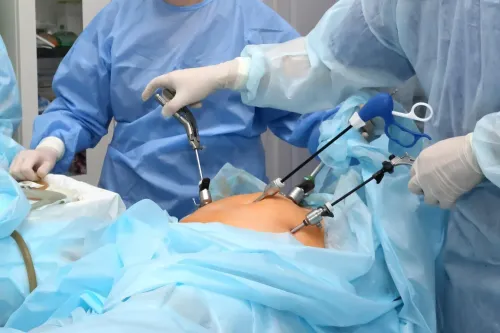
- Arrange urgent surgery for an abscess, infected joint, deep tissue infection, extensive dead tissue, or infection that cannot be controlled with outpatient care.
- Consider hospital treatment for severe infection and for selected moderate infections with rapid progression, major comorbidities, poor oral-drug absorption, or inadequate home support.
Absent pulses, gangrene, or suspected ischaemia require a vascular referral because poor blood flow prevents healing; infection plus ischaemia needs urgent specialist coordination. Dr. Shaunak Saha can help assess whether surgical management or referral is appropriate when a wound is deteriorating.
Frequently asked questions
Why can a small diabetic foot wound become dangerous?
Neuropathy can reduce protective sensation, so pressure, heat, a foreign object, or a deepening wound may go unnoticed. Poor blood flow and high blood glucose can also slow healing and allow infection to spread.
Which diabetic foot wound warning signs need same-day medical attention?
Seek same-day care for spreading redness, warmth, swelling, pus, foul odour, fever, chills, red streaks, black or grey skin, exposed bone, rapidly worsening swelling, or a new deep wound.
How do clinicians determine how deep a diabetic foot wound is?
They inspect the wound’s size, edges, drainage, colour, surrounding skin, and depth. They may gently probe the wound to check whether it reaches tendon, joint, or bone, while assessing sensation and circulation.
Which tests can reveal infection, bone involvement, or poor blood flow?
Blood tests can show inflammation and infection. An X-ray can detect gas, a foreign body, or later bone changes; MRI gives more detailed images when bone infection is suspected. An ankle-brachial index, toe pressure, or Doppler ultrasound evaluates blood flow.
What early treatment helps prevent a diabetic foot wound from worsening?
Treatment can include cleaning and dressing the wound, removing dead tissue, taking pressure off with suitable footwear or a cast, treating infection with prescribed antibiotics, and improving blood flow when circulation is inadequate.